We preserve your precious teeth without extraction.
- My gums are swollen and pus is coming out.
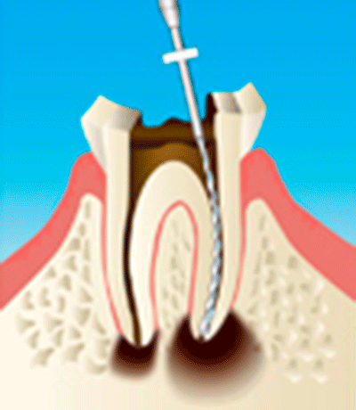
- I had root canal treatment, but for some reason the pain and swelling won't go away
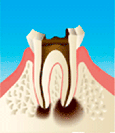
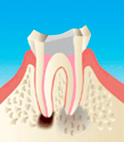
- I was told that the bone supporting my teeth is dissolving.
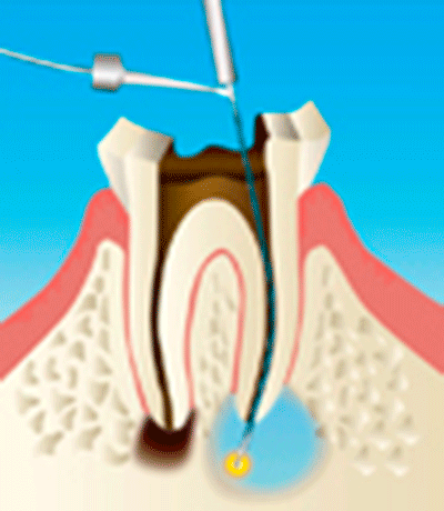
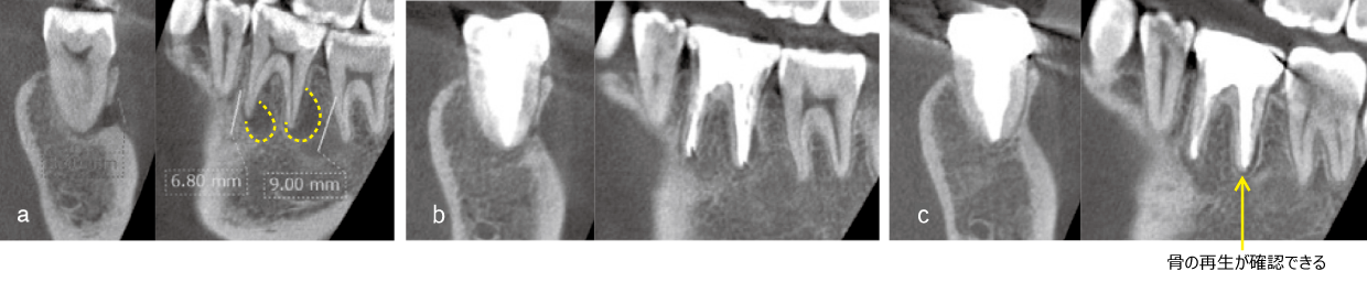
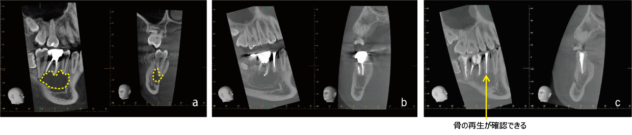
EMAT is a regenerative treatment that revives the "alveolar bone" that supports teeth by activating bone-forming cells called "osteoblasts," which are naturally present in the patient. At the same time, it has the effect of sterilizing bacteria that inhibit the preservation of teeth, so it can be expected to improve the symptoms you are suffering from.
In order to respond to everyone's desire to "heal without extracting teeth," ElectroMagnetic Apical Treatment(EMAT) was born as a result of many years of research.
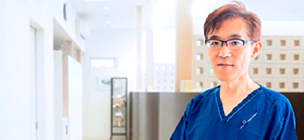 We are the only clinic run by Toshihiko Tominaga, the developer of EMAT treatment.
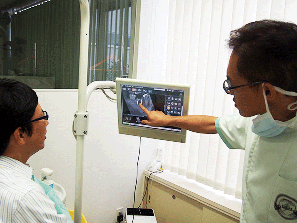
 We use specialized diagnostic methods and treatment methods based on our past experience.